NEW Inverted microscope system
IXPLORE IX85
Clearer Insights,More Discovery
The IXplore™ IX85 platform delivers an unmatched level of customizability, allowing you to design or build an intelligent, high-performance imaging system that meets your specific goals.
And with an industry-leading field number (FN) of 26.5mm plus an array of advanced end-to-end imaging and workflow features, the IXplore™ IX85 means you can capture more than ever before while dramatically reducing your acquisition times.
Expand Your View, Enhance Your Vision
See and capture more—and reduce your acquisition times—with an unmatched field of view (FOV) in the industry and an array of advanced end-to-end imaging features that set a new standard in clarity and precision, including groundbreaking new objective technology
Unmatched 26.5mm FOV
View more of your sample at once with an industry-leading field number (FN) of 26.5mm on two integrated imaging ports. Cover larger areas in fewer images, get more data in each image, minimize the need for image stitching, and speed up your workflows.
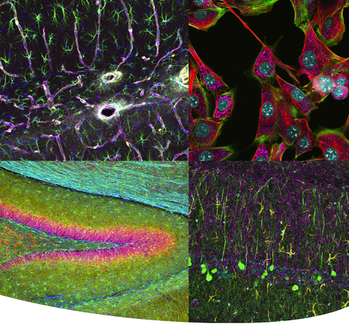
Cultured NIH 3T3 cells. Blue: nuclei, Green: tubulin, Red: HSP60, Gray: fibrillarin. Sample provided by EnCor Biotechnology Inc.
Experience New Levels of Precision Clarity
With an innovative, strategic selection of advanced beginning-to-end imaging features, the IXplore™ IX85 is setting a new standard in precision and clarity—enabling you to see more fine details than ever before and easily obtain impactful, publication-ready images.
See Deeper into Your Samples with Our New Multi-Immersion Objective
Our new multi-immersion objective (LUPLAPO25XS) with groundbreaking new immersion technology lets you see deeper into your samples and reveal structures that were previously out of reach.
New Experience with Silicone Gel Pad
LUPLAPO25XS
NA0.85 / WD 2.00mm*
Immersion: Silicone gel pad, Silicone oil, Water
* approx. 1mm for silicone gel pad
Automated Acquisition Features
The IXplore™ IX85’s automated acquisition features, customizable interfaces, and efficient task management software allow you to work more efficiently while staying confident in your results. Your productivity is further enhanced by advanced real-time image processing and analysis.
Time-saving innovations include the pioneering new gel objective, a new auto correction collar, AI-based automated sample finding, TruAI™ Z-drift compensator, effortless contrast control, and simplified well-plate acquisition—all backed by customizable interfaces that let you design your system to the way you work best.
TruAI™ Z-drift compensator
Your sample always in focus
Near-Infrared Imaging Solutions
Near-Infrared Imaging Solutions
See in red—open a spectrum of possibilities. When investigating cellular
processes in the context of their 3D environments, model organisms and
organoids are often superior to 2D culture systems. Near-infrared (NIR)
microscopy has proven beneficial for imaging biological tissues due to a
lower absorption and scattering of NIR light than visible light.
TIRF Imaging
Designed for membrane dynamics, single molecule detection, and colocalization experiments, the IXplore TIRF microscope system offers simultaneous multicolor TIRF imaging
TIRF Objectives
Total internal reflection fluorescence (TIRF) is facilitated by a wide range of objectives featuring a high signal-to-noise ratio and a correction collar to adjust for cover glass thickness and temperature.
Our corrected plan apochromat objectives with an NA of 1.5 help you acquire uniform high-quality images with a large field of view. Take advantage of Olympus' remarkableTIRF objective with the world’s highest NA, 1.7*.


