ArcturusXT™ Microdissection System
![]() Flexible and Modular Laser Capture Microdissection
Flexible and Modular Laser Capture Microdissection
- Laser Capture Microdissection and UV Laser Cutting
- Open, Modular Platform
- Simple, Intuitive Operation
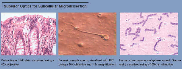
- Superior Image Quality
- Sample Custody Maintained at all Times
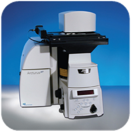 |
The ArcturusXT™ system from MDS Analytical Technologies is a unique microdissection instrument that combines Laser Capture Microdissection (LCM) and Ultraviolet (UV) Laser Cutting in one platform. The open, modular design of the ArcturusXT system provides unparalleled research flexibility and versatility. The system removes the guesswork from the microdissection process by allowing researchers to maintain custody of the sample throughout the experiment, ensuring that only the desired material has been collected. |
The solid-state IR laser, exclusive to MDS Analytical Technologies’ microdissection, delivers a gentle capture technique, which preserves biomolecular integrity and is ideal for single cells and small numbers of cells. The solid-state UV laser delivers unprecedented speed and precision, well-suited for microdissecting dense tissue structures and for capturing large numbers of cells.
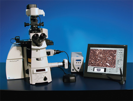
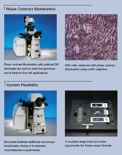
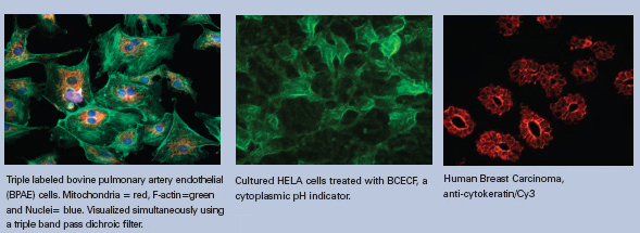
The ArcturusXT microdissection instrument is modularized to suit any dynamic research need. The system utilizes a Nikon Eclipse Ti-E inverted research microscope with a variety of options available at the time of purchase or post-purchase. Each instrument has IR-enabled LCM, an interactive pen-display monitor, and a trackball actuated stage for easy and ergonomic navigation. The system may be configured with LED brightfield illumination or high-intensity halogen illumination, enabling Phase Contrast and Differential Interference Contrast (DIC) microscopy applications. The ArcturusXT system may be purchased with or without UV laser cutting and attenuable fluorescence. A wide range of objectives are available, including 2X–100X Dry, and 100X Oil (see back page for complete list). For ultimate image quality and analysis, a high-resolution megapixel camera may be added, which includes the MetaVue® imaging system. A second flatscreen monitor may be included for maximum workstation efficiency.
Harvest single cells or tens of thousands of cells in seconds.
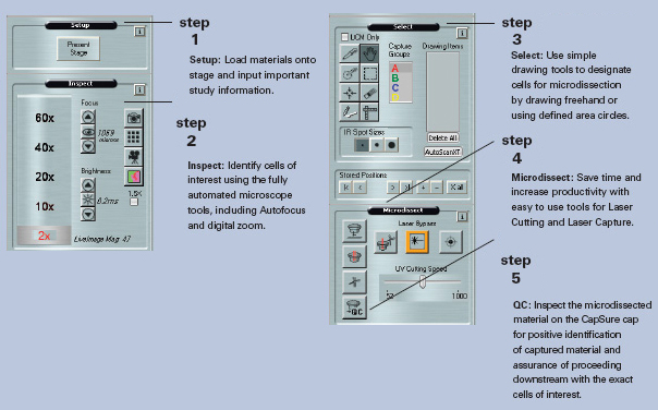
The simple, intuitive user interface places the focus on research. From sample loading to extraction of biomolecules, the uncomplicated workflow maximizes experimental efficiency.
Automatic electronic documentation may be employed to record each step of the process— before and after microdissection. Static images and live video can be taken at any point during the process, providing a record of the entire experiment. View the CapSure® LCM cap at the QC station for positive identification of captured material, and utilize Capture Groups to display and track all individual and group area measurements
The AutoScanXT image analysis software module automatically identifies cells and regions based on user-defined criteria, greatly reducing the overall time required to perform a microdissection experiment. This optional module performs optimally on high-contrast samples and may be used with colorimetric, fluorescence, and IHCstained specimens. Once the regions have been automatically identified, the user proceeds directly to standard microdissection using the ArcturusXT instrument.
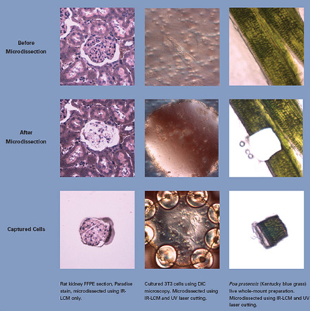
The unique combination of IR laser capture and UV laser cutting permits the use of any slide type and any sample preparation. Choose from glass, glass membrane or framed membrane slides for contact or non-contact microdissection. Any specimen preparation may be used:
- thin or thick sections
- frozen or formalin-fixed tissues
- chromogenic stained, fluorescently stained, or unstained sections
- hydrated or dehydrated specimens
- fine needle aspirates
- forensic smears
- live plant whole-mount preparations
- cell cultures
The ArcturusXT system’s open platform can be upgraded and expanded to meet changing research requirements. The available microscope port allows users to modify the system for alternate applications, such as adding a second camera for high-resolution imaging. The open system design also makes possible the easy exchange of stage inserts to accommodate future assay formats and can be equipped with an environmental control chamber, providing optimal conditions for live cell and cell culture applications.
LRI Instrument employs a team of highly qualified and expertly trained application scientists and technical support specialists to assist researchers in any research area.
Technical Specifications
Performance Specifications
Stage: |
Motorized with positions for three slides and four caps, trackball actuated in X and Y axes with 1 μm precision |
Objectives: |
Motorized turret with 2X, 10X, and 40X Nikon objectives (optional 4X, 20X, 60X, 100X Dry and 100X Oil Nikon objectives available) |
Filter: |
Six-position fluorescence filter turret with three filter cubes and three available positions for application-specific filter cubes |
Color |
Excitation |
Emission |
Red |
570–630 nm |
> 655 nm |
Green |
503–548 nm |
> 565 nm |
Blue |
455–495 nm |
> 510 nm |
UV (optional) |
340–390 nm |
> 410 nm |
Triple Dichroic (optional) |
||
DAPI |
385-400 |
450-465 |
FITC |
475-493 |
503-533 |
TRITC |
545-565 |
582-622 |
Epi-Fluorescence: |
2000-hour broad-spectrum Metal Halide lamp; userreplaceable with no alignment required |
Brightfield: |
100 W halogen or hig-hintensity LED illumination system |
Lasers: |
Solid-state, near-IR laser (810 nm) |
Camera: |
1024 x 768 1/3" color CCD with electronic shutter to 1/10,000s and on-chip integration to 30 seconds |
Altitude: |
For use up to 6600 ft. (2000 m) |
Operating temp.: |
18–30°C |
Operating humidity: |
≤ 60% relative humidity (non-condensing) |
Dimensions: |
30" (D) x 22" (W) x 29" (H). |
Power: : |
100–240 Vac, 50–60Hz; 600 W |
Work surface req.: |
36" x 72" (92 cm x 183 cm) |
Computer: |
2.8 GHz Pentium® 4 (min.), 2 GB RAM (min.), 80 GB hard drive (min.), DVD +/- RW drive, Windows® XP Pro, 1280 x 1024 dpi resolution monitor, and interactive pen display monitor |
- Stage translation
- Slide selection
- Focus and light intensity
- Laser parameters
- Objective selection
- Cap transfers (including QC confirmation)
- Camera settings
Image file format: |
May be captured, annotated, and saved as JPEG or TIFF files |
